Bart Smeets


About me
At the head of the Multicellular System Dynamics Lab at KU Leuven, I work at the interface of active matter physics and mechanobiology. At our lab, we love building computational models of multicellular systems. For this, we mainly develop agent-based models focusing on mechanics that represent interaction between cells and subcellular elements. We have been spearheading the development of detailed foam-like descriptions of tissues. These models are applied to various biological and technological processes, including embryonal development, tumor growth, in biofabrication operations (such as 3D printing), or in bacterial biofilms. Please also have a look at our lab's Youtube page!
Publications
Click here for a complete list of publications, or look me up on Google Scholar. Below, I discuss some important and/or recent work from our group.Highlighted publications and preprints
Biomechanics and Modeling in Mechanobiology (2026): Homeostatic pressure of a proliferating multicellular foam with hydromechanical volume regulation
Jef Vangheel, Jeroen Guillierme, Irish Senthilkumar, Enda Howley, Eoin McEvoy & Bart Smeets
How do cell-scale mechanics determine how tissues grow and outcompete each other? In this work, we develop a hydromechanical active foam model that couples two key ingredients: i) a foam-based mechanical description of multicellular aggregates and a ii) biophysical model of osmotic cell volume regulation. Individual cells are modeled as proliferating, pressurized bubbles whose growth emerges from ion-water transport, cortical mechanics and interfacial tensions, allowing tissue-scale mechanics to arise directly from single-cell physics.
The model predicts that growth in confinement naturally reaches a hydromechanical homeostatic state characterized by a well-defined homeostatic pressure, generated without imposing phenomenological growth laws. A central prediction is that decreasing effective interfacial tension (either by lowering cortical surface tension (softer cells) or by increasing net adhesive tension between cells) raises this homeostatic pressure at the tissue scale. This gives softer and more adhesive cell populations a mechanical competitive advantage, allowing them to displace even faster-growing but mechanically disadvantaged populations. This study shows how multicellular growth can be understood as a hydromechanical instability governed by interfacial mechanics, pressure generation and volume checkpoints, yielding testable predictions for tissue competition and tumor mechanics.
PNAS (2026) (BioRxiv preprint): Stress-dependent growth of breast cancer models arises from a cellular volume checkpoint
Irish Senthilkumar, Jef Vangheel, Vatsal Kumar, Laoise McNamara, Bart Smeets, Enda Howley, Eoin McEvoy
The growth of a tumor generates a mechanical stress which is transmitted to its surrounding extracellular environment. In this collaboration with the lab of Eoin McEvoy from the University of Galway, we investigate the stress-dependent growth of breast cancer spheroids that are growing in an elastic environment. For this, we use a biophysical model for cell growth governed by actively controlled osmolarity, which is integrated in a computational foam model of tissue growth.
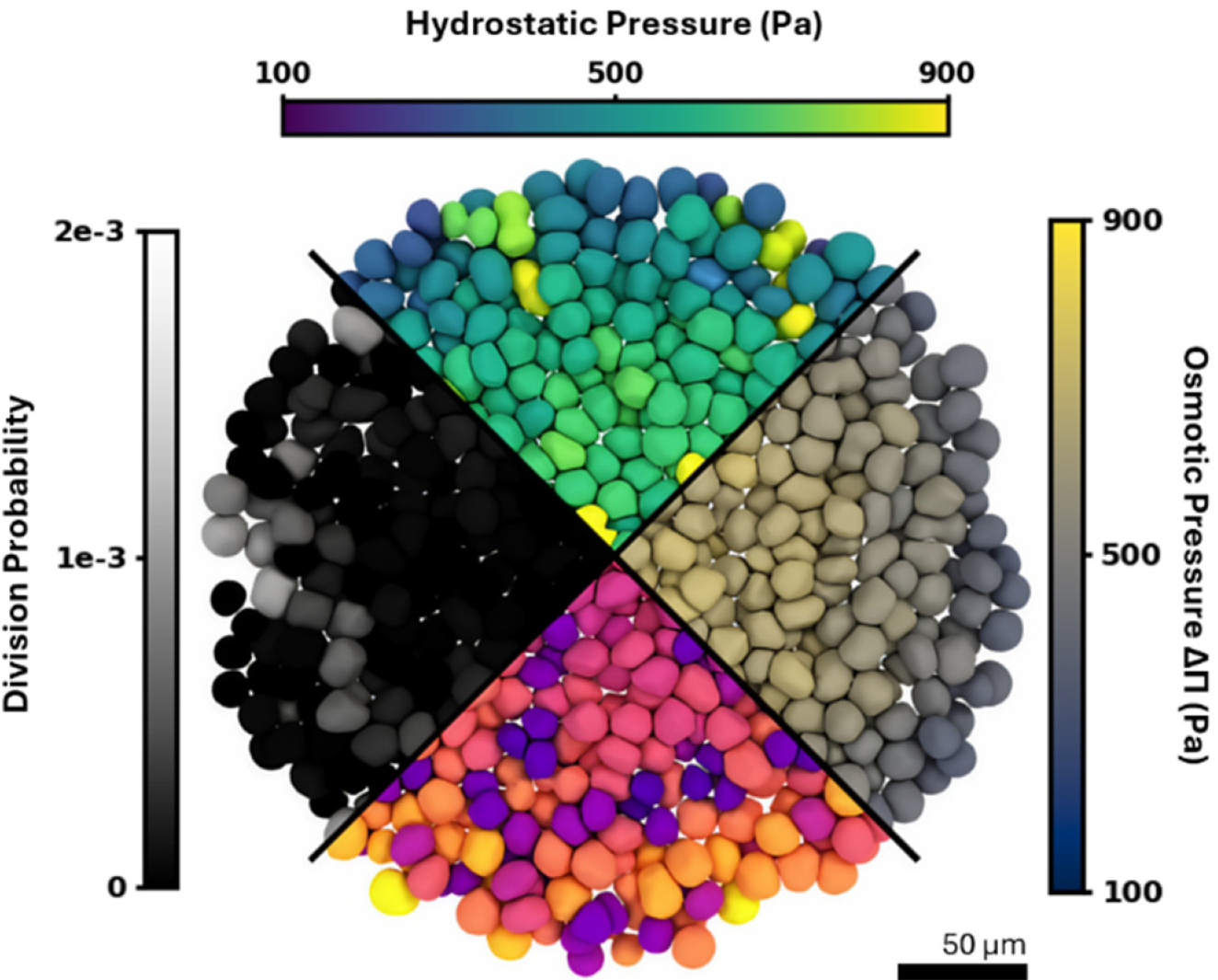
The mechanical behavior of the extracellular environment is approximated as a neo-Hookean elastic material. Simulation of the material deformation in function of internally generated pressure (the growth pressure generated by the spheroid) can be done using the Finite Element Method. However, the computational model evaluates the growth across more than 12 days, and the hydromechanical foam model uses small time steps to accurately simulate water and ion flux in the cell. At these timescales, the incremental deformation of the extracellular environment is very small. However, the Finite Element solution of this problem is very costly. To render feasible simulations, we thus use an AI-supported framework, where we trained a neural network model to predict the boundary displacement based on input forces. This drastically reduces the computational cost of the elastic response of the environment, which, while mechanically very important, is computationally rather 'boring'. Using this framework, our model is able to simulate the growth dynamics of experimental cancer spheroids in environments of different stiffness. This study demonstrates how new AI-based approaches do not replace but support mechanistic models, helping them focus on the scientifically relevant or novel parts of the simulation, in this case the hydromechanical regulation of tumor size.
Physical Review Research (2026) (BioRxiv Preprint): Rigidity transitions in a 3D active foam model of cell monolayers with frictional contact interactions
Vangheel, J., Ramon, H. and Smeets, B.
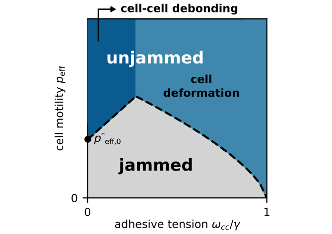
The classic vertex model predicts a density-independent rigidity transition to occur when preferred cell shape exceeds a critical shape index. In other words, factors that drive cell elongation, such as adhesion / reduced interfacial tension will help unjam the tissue. This prediction is in marked contrast with the behavior of colloid-like (active) particles. For these particles, adhesion will inhibit particle rearrangments and thus drive the system towards a glass or jamming transition.
We developed a 3D foam-like model of cell monolayers in which cells are represented as discrete entities with explicit contact interactions. This model naturally represents foam-like behavior (where the microstructure is determined by the balance of tension at various interfaces): At high cell-cell adhesion (or reduced interfacial tension at the cell-cell interface), cells become columnar and can even extrude from the monolayer. At high cell-substrate adhesion, cells become spread out and squamous. The foam was turned into an 'active foam' by including a minimal model of cell motility, which includes both a polar component (net migration) and a dipolar component (cell elongation). Our model captures the essential features of both vertex models (fluidity driven by cell deformation) and colloid-like models (fluidity driven by debonding). Thus, we use the model to draw a unified phase diagram of rigidity transitions in foam-like materials. Moreover, we were able to explore the effect of cell-cell friction. We show that it drastically reduces cell rearrangements (thus appearing to solidify the tissue). However, this was not found to be a (typical) rigidity transition, since the static yield stress in a shear experiment remains zero, even when increasing cell-cell friction.
Nature Communications (2025): Emergence of bidirectional cell laning from collective contact guidance
Ongenae, S., Svitina, H., Belpaire, T.E., Vangheel, J., Martens, T., Vanden Berghe, P., Papantoniou, I. and Smeets, B.
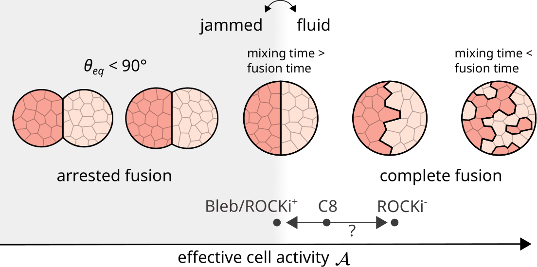
It has long been known that multicellular systems may exhibit liquid-like behavior. For example, tissues will wet or dewet from a surface or phase separate during cell sorting. When brought together, two spheroids (spherical droplets of cell material) will spontaneously fuse, much like liquid droplets with surface tension. Yet, when probed by mechanical tests, tissues appear solid-like. These liquid-like behaviors stem from active cell forces driving rearrangements and cytoskeletal remodelling, dominating the long-timescale viscous response of tissue.
In this work, we studied the fusion of tissue spheroids to investigate how active properties at the cell scale influence macroscopic fusion dynamics. We performed a detailed experimental analysis of both the fusion process and tissue microstructure at the cellular scale. We found that cells in fusing tissues are highly active, with frequent rearrangements. We then compared these experiments to simulations of an active foam model, where cells are modeled as volume-preserving 'bubbles' with surface and adhesive tension. Migration was modeled via a protrusive/retractive pressure aligned with a randomly diffusing polarity. These simulated spheroids spontaneously fused — just like in experiments. We mapped how cell-scale properties govern the outcome of fusion, and propose a dimensionless activity parameter that predicts whether the tissue behaves fluid- or solid-like, and whether fusion succeeds.
Nature Physics (2024): Emergence of bidirectional cell laning from collective contact guidance
Lacroix, M., Smeets, B., Blanch-Mercader, C., Bell, S., Giuglaris, C., Chen, H.Y., Prost, J. and Silberzan, P.
In this work, we investigate a remarkable finding: When let HBEC cells migrate on micropatterned grooves, they spontaneously form very large collective streams or 'lanes' that align with the direction of the groove. These streams have no preferred direction, so streams of opposite direction appear and compete with each other, leading to a gradual coarsening into stable, bidrectional lanes. We use a combination of active hydrodynamic theory (work of Carles Blanch-Mercader) and particle-based simulations to explain the emergence of these lanes. We find that micropatterned grooves can be thought of as an aligning field, for example through anisotropic friction, which is lower in the direction of the grooves. This triggers a disordered-to-polar phase transition that stabilizes into large polar lanes. Both simulations and hydrodynamic theory predict a phase diagram of collective streams with varying anisotropy ratio (in the experiments modulated by groove depth) and flocking strength.
The flocking strength is based on the existence of a velocity-polarity coupling: As neighbours influence a cell's velocity, the cell starts to reorient into its changed velocity. Thus, if this coupling is sufficiently strong, mechanical interactions (which can be purely repulsive) between adjacent cells drive a disordered-to-polar phase transition. While not observed in experiments, even without any grooves the disordered-to-polar phase transition is expected to occur at high enough flocking strength. Below you see a movie of a very large simulation of a polar fluid of self-propelled particles at high flocking strength. With time, polar domains grow ever larger and very large collective streams, spanning thousands of particles, appear. Since there is no global aligning field in this case, there is no prefered direction in the collective movements.
Biophysical Journal (2023): Stability of asymmetric cell division: A deformable cell model of cytokinesis applied to C. elegans
Cuvelier, M., Vangheel, J., Thiels, W., Ramon, H., Jelier, R. and Smeets, B.
The nematode C. Elegans is a model organism to study embryonic development. In contrast to higher organisms, the fate of every cell is deterministic, and the full development of every cell in the organism has been fully characterized. In early development, various symmetry breaking events happen that create complexity in the organisms, typically through a sequence of asymmetric cell divisions. These asymmetric divisions not only play a intracellular biological role (with different materials and building blocks available for different daughter halves), they also have a pronounced effect on morphogenetic patterning.
In this work, we introduced for the first time a model for asymmetric cell division to a foam description of multicellular tissues. In this model, cytokinesis is triggered by the action of a contracting furrow ring. By placing the ring at an offset from the midplane, and an internal stiffness that prevents collapse from balloon instability, the cell will divide into two daughter cells with different volumes. Taking data of division planes and division offsets from experiments, we thus simulate the early development of the embryo. Up to the 6 cell stage, this info alone, supplemented with basic foam-like behavior, is enough to predict the topology, positions and relative volumes of different cells. This study is a proof-of-concept for the use of foam-like descriptions that include proliferation to help understand morphogenetic processes.
Research
Modeling cells as foams
The idea of comparing tissues to foams goes back to the classic work of D’Arcy Thompson. He noticed that the spatial organization of multicellular systems—animal embryos, plant tissues, or simple multicellular organisms—often resembles the packing of bubbles in a foam. This analogy is not only visually appealing; it is also powerful from a mechanical point of view. In a foam, the mechanical behavior of each bubble is governed by surface tension and internal pressure. A similar idea works surprisingly well for cells. Many mechanical features of a cell can be captured by an effective surface tension at its boundary and an internal pressure generated by the cytoskeleton and osmotic forces. With this representation, geometry and mechanics become directly linked at the level of individual cells. The Young–Laplace law connects surface tension and pressure difference to the curvature of a cell’s boundary, while contact angles at junctions reveal the relative tensions between neighboring cells or between a cell and a substrate. This “cell as a bubble” view is also motivated by cell architecture. Most cells have a thin, contractile cortex beneath the membrane that functions like a surface under tension, together with a more or less uniform pressure inside. These ingredients make the foam analogy natural and useful.
Of course, biological tissues are not passive foams like those in a soap bath. Cells are active. They continuously adjust their interfacial tensions in response to biochemical signals, cell shape, and mechanical forces. Many cells also have built-in asymmetries, such as the apico–basal polarity of epithelial cells. They can migrate, extend protrusions, retract, divide, and grow. Cell division, for example, involves an active process that splits one “bubble” into two. Because of these active processes, a tissue is better viewed as an active foam. Studying tissues through the lens of active foams allows us to understand how local mechanical rules—tension, pressure, adhesion, and activity—shape global tissue behavior. This approach helps explain how tissues rearrange during development, how they flow or solidify, and how they transition between fluid-like and solid-like states. In this way, the foam model provides a simple language for describing the complex and dynamic material properties of living tissues.
Contact
Email: bart.smeets@kuleuven.be
Adress: Kasteelpark Arenberg 30, 3001 Heverlee, Leuven, Belgium
Contact me if you are interested in our research! I am always looking for good students or interesting new collaborations!